Ledipasvir-Sofosbuvir Can Improve HCV-Mediated Neurocognitive Dysfunction
A late afternoon talk on the third day at The Liver Meeting 2014, evaluated the influence of some of the newer antiviral agents on "brain fog," a phenomenon quite commonly observed in hepatitis C virus-infected patients, especially among those with mild disease.
A late afternoon talk on the third day at The Liver Meeting 2014, an annual event by the American Association for the Study of Liver Disease, held in Boston, Massachusetts, evaluated the influence of some of the newer antiviral agents on “brain fog,” a phenomenon quite commonly observed in hepatitis C virus (HCV)-infected patients, especially among those with mild disease.1
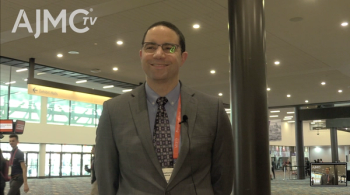
The talk was entitled, “Cerebral MR spectroscopy and patient-reported mental health outcomes in hepatitis C genotype 1-naïve patients treated with ledipasvir and sofosbuvir.” Nezam H. Afdhal, MD, director of hepatology at Beth Israel Deaconess Medical Center and associate professor of medicine at Harvard Medical School, who presented the data, explained that HCV-infected patients often have difficulty focusing, functioning, and with neurocognitive performance. “The fact that the virus is detected in the subcortical white matter and the brainstem might explain the occurrence,” he said.
According to the authors, changes associated with HCV infection result in increases in magnetic resonance spectroscopy (MRS)-measured myoinisitol (MI) and choline (CH), and reduced n-acetyl aspartate (NAA). So the authors hypothesized that viral suppression with ledipasvir/sofosbuvir (LDV/SOF) ± ribavirin (RBV) could influence neuronal function, as measured by MRS, and could be correlated with mental health constructs of patient-reported outcomes (PROs). This study was piggy-backed onto the ION-1 trial—HCV treatment-naïve patients with F0-F2 fibrosis were enrolled from a single trial site. MRS analyzed signals from CH, creatine (Cr), NAA, and MI from basal ganglia, frontal, and dorsolateral prefrontal regions at baseline, week 4 post-treatment, and week 12 post-treatment—periods when the sustained virological response (SVR) is measured. The 3 biomarkers—MI, CH, and NAA—were quantified by normalizing against Cr, and PROs were determined at the same time points using validated questionnaires, as was alanine aminotransferase and the viral load.
The study found a significant increase in NAA/Cr in the basal ganglia at SVR12, suggesting an improved neuronal function. Better outcomes were observed in RBV-free regimens. While SVR4 showed a tendency for improvement, the NAA/Cr ratio did not demonstrate a statistically significant increase. However, a significant decrease in CBF was observed after 4 weeks of treatment, and recovery was observed after 12 weeks. Based on these observations, the authors concluded that the NAA/Cr ratio could be an adequate marker of neuronal recovery following therapy. “Changes in the metabolite pattern captured by MRS may be associated with changes in PROs related to mental health,” said Dr Afdhal. He drew attention to the fact that their results with the RBV-containing regimen indicate impaired neuronal function and recovery. Dr Afdhal revealed that following this exploratory trial, a fully-powered placebo-controlled study is ongoing.
References
- Byrnes V, Miller A, Lowry D, et al. Effects of anti-viral therapy and HCV clearance on cerebral metabolism and cognition. J Hepatol. 2012;56(3):549-556.
The authors conducted arterial spin labeling and cerebral blood flow (CBF) MRI in 14 patients, 8 of whom were male. The patients were relatively young, with a high viral load (>800,000 IU), but mild disease (
Newsletter
Stay ahead of policy, cost, and value—subscribe to AJMC for expert insights at the intersection of clinical care and health economics.
