Genetics of Osteogenesis Imperfecta and Phenotypic Implications
Osteogenesis imperfecta, a skeletal disorder caused by mutations in type I collagen, exhibits structural and functional differences on a genotypic level that contribute to diverse phenotypes.
Osteogenesis imperfecta (OI), a skeletal disorder caused by mutations in type I collagen, exhibits structural and functional differences on a genotypic level that contribute to diverse phenotypes.
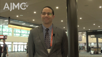
Brendan Lee, MD, PhD, from the Baylor College of Medicine discussed the genetics of OI and homeostatic mechanisms in the skeleton at the 2014 American Society of Bone and Mineral Research Conference in Houston, Texas.
Human genetics of the skeleton can be analyzed in multiple ways. Scientists can examine phenotype (for example, radiologic evidence in skeletal morphology and bone density), candidate genes (including essential transcription factors and their targets), signaling pathways, and phenotypic expansion (new alleles). Once they understand the genetic underpinnings of skeletal disease, scientists may be able to develop targeted therapy to address diverse phenotypes.
Dr Lee’s lab is examining bone matrix, mineralization, and density in OI. OI ranges in debility from increased vulnerability to fractures to severe disease involving deformity and contractures.
In OI, patients suffer from low bone mass, bone fragility/deformity/fractures, and extra-skeletal manifestations. They exhibit a wide phenotypic spectrum. For example, some OI only involves bone disease, while Bruck syndrome is a type of OI with significant contractures.
This variation brings up questions of how structure and function on a genotypic level contribute to the diverse phenotypes. In one experiment, Dr Lee evaluated the differential contribution of chaperoning. He found that mice deficient in prolyl 3-hydroxylase 1 (P3H1) show decreased trabecular bone mass as well as ligament, tendon, and skin manifestations, suggesting that hydroxylation of this protein is required in tissue.
Brittle bone disease can be caused by abnormalities in cellular trafficking, matrix cell signaling, or matrix collagen cross-linking. In another experiment using a mouse model of OI, Dr Lee studied the overlap of the CRTAP phenotype with increased TGF-beta signaling. He found decreased bone mass, increased bone turnover, and increased osteocyte density. Inhibition of TGF-beta improved trabecular bone mass in the spine, suggesting that anti-TGF-beta may be used as a treatment modality.
Dr Lee said, we now “see a whole spectrum of rare mutations underscoring different mechanisms of disease.” He then described a third experiment that showed that semi-dominant WNT1 mutations were present in early onset osteoporosis and in OI as a dose-response requirement of a very dominant phenotype.
He concluded by noting that we now have a wide variety of treatments for OI, including bisphosphonates for pediatric OI, teriparatide for adult OI with quantitative defects of collagen, and anti-TGF-beta for various other types of OI.
Dr Lee said that there isn’t “one simple thing that explains it” for OI abnormalities. However, just like there is an overlap between OI and osteoporosis, there is a question of how genetic variants contribute to osteoporosis. Individualized treatment of osteoporosis may be the next important step in therapeutic intervention.
Newsletter
Stay ahead of policy, cost, and value—subscribe to AJMC for expert insights at the intersection of clinical care and health economics.


